|
magnified
scene by clicking image
| Fig.1 |
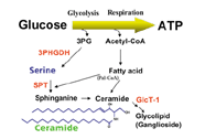
Glucose metabolism in brain |
magnified
scene by clicking image
| Fig.2 |
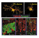
(Upper)
Influence of serine in medium on cultured Purkinje cells; (lower) expression pattern
of 3PHGDH, a key enzyme for serine biosynthesis in cerebellar tissues. |
|
Why
study serine?
In general, once
neurons are differentiated, they lose the ability for roliferation and instead
survive until the death of the organism. Accordingly, in thecase of humans, neurons
continue to survive and perform neurological activities for more than 80 years.
That neurons have such exceptional power is amazing and we believe that serine
is one of the key valuables in understanding how neurons survive for such long
periods.
If you were asked which compounds were indispensable for neurons to survive, you
would probably readily list glucose and oxygen. Afterall, neurons completely depend
on adenosine triphosphate (ATP) in order to perform neuronal activities, and glucose
and oxygen are used mainly to produce ATP. However, approximately 30% of glucose
is used for purposes other than ATP production (Fig. 1). Our laboratory carried
out a study that aimed to clarify the molecular basis of the biological potential
of neurons, focusing on serine, which is synthesized from 3-phosphoglycerate,
an intermediate in glycolysis. At the end of our study, we concluded that serine,
which is a common compound classified in the nonessential amino acid group, is
essential for development, survival and morphogenesis of neurons. To our surprise,
synthesis of serine from glucose is mainly carried out in astrocytes, which are
neuroglial cells. Why is this amino acid so important for neurons? We propose
that sphingolipids, which are synthesized from serine, are the key to answering
the above question. Our study shows that there is dynamic interaction between
neurons and neuroglia cells, which can be observed through the metabolism of serine,
and that this is the key to understanding the reason why neurons can live for
such an extended period of time.
Uniqueness
of serine metabolism in the brain
In 1998, we found
that hippocampal neurons cultured in a medium without serine or glycine resulted
in poor morphological development of cells and a rapid decrease in the number
of surviving cells. Serine is required not only in the hippocampal neurons, but
also in Purkinje cells. However, such neurotrophic activity was not observed for
D-serine, which is an optical isomer of serine and is specific to both serine
and glycine. In order to understand why serine supports survival maintenance,
3PHGDH expression, which is a key enzyme in the synthesis of serine, was analyzed.
As shown in Fig. 2, the 3PHGDH expression level was extremely low in neurons,
while that in surrounding astrocytes was markedly high. This result suggests that
neurons themselves do not have the ability to synthesize serine and that neurons
completely depend on neuroglial cells for their serine supply. Clarification of
the mechanism underlying the cell-specific expression of the 3PHGDH gene in the
brain is expected in the future.
magnified
scene by clicking image
| Fig.3 |
Generation
of knockout mice with tissue-specific deficiency using a Cre/LoxP recombination
system |
magnified
scene by clicking image
| Fig.4 |
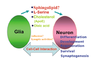
Reciprocal
interaction between neurons and neuroglial cells, which regulates the processes
of differentiation and morphogenesis of neurons, and the formation of synapses.
The compounds that mediate the interaction are those which are commonly known. |
|
Signaling
and microdomains formation by sphingolipids derived from serine
Why is serine
particularly important for the survival of neurons? Serine released from neuroglia
cells may be transported to neurons via a transporter for a neutral amino acid,
and used for the synthesis of various biomolecules. We are now focusing our attention
on sphingolipids present on the cell membrane. Major sphingolipids, such as sphingomyelin
and gangliosides, are abundant in the brain. The sphingolipid biosynthesis starts
from a condensation reaction between serine and palmitoyl CoA catalyzed by serine-palmitoyl
transferase (SPT). Accordingly, a shortage of serine in neurons leads to a decrease
in the cellular content of sphingolipids, and finally leads to neuronal death.
Therefore, we had to ask how neuronal death is induced by the decrease of sphingolipids
content.
It is generally considered that sphingolipids have two unique functions: one is
that sphingolipids themselves are signalling compounds associated with the survival
and death of cells, and the other is that sphingolipids form aggregates in the
presence of cholesterol and GPI-anchored glycoproteins, and form microdomains,
called rafts, on the cell membrane. Rafts are considered to play an important
role as a base for receiving and sending information inside and outside of the
cell, or as a processing base for the amyloid protein. Therefore, a decrease of
sphingolipids, which play such an important role, may have a significant influence
on neurons such that their absence threatens the survival of neurons. Further
studies, however, are needed to clarify the precise mechanisms underlying neuronal
death.
Analysis
of functions in a multicellular system: generation of knockout mice with tissue-specific
manner
Do serine and
sphingolipids function as survival factors for neurons in actual living organisms?
To answer this question, using a generation of 3PHGDH- and SPT- (genes encoded
with key enzymes related to the syntheses of serine and sphingolipids, respectively)
knockout mice is the most reliable method. Since it is possible that both 3PHGDH
and SPT are lethal genes at the embryonic stage, we adopted a method for generating
conditional knockout mice (Fig. 3). Thus far, we have succeeded in generating
skin- and T-cell-specific sphingolipid-deficient mice; as a result, analysis of
the functions of sphingolipids in various tissues is possible.
Recently, a very interesting study related to the functions of lipids in the brain
was reported in Science. The study indicated that cholesterol/apo E, released
from neuroglial cells, is extremely important in the formation and maintenance
of synapses. Thus the following question arises: Are sphingolipids released from
neuroglia cells and do they send signals to neurons or, are there any factors
released from neurons influence the activity and function of neuroglial cells
(Fig. 4)? We are expecting that our knockout mice will provide essential information
that will clarify the functions of serine and lipids inside and outside of neurons.
Conclusion
Since the human genome sequence was completed, the identification of causal genes
of hereditary diseases is being carried out at a rapid pace. A serine-deficiency
disease (a case of West syndrome) was discovered to be caused by 3PHGDH gene mutation,
and hereditary sensory neuropathy (HSN-1) by mutation of the gene encoding SPT.
These diseases clearly show the importance of serine metabolism in the human.
We expect that progress in brain research, including research not only on neurons
but also on neuroglial cells, will deepen our understanding of the brain, as well
as provide measures to prevent and cure various degenerative brain diseases and
maintain or protect the functions of the human brain.
The authors would like to thank the many people presented in the reference section
for their research.
<References>
1) Mitoma, J., Furuya, S., and Hirabayashi, Y.: A novel metabolic communication
between neurons and astrocytes: A non-essential amino acid L-serine released by
astrocytes is essential for developing hippocampal neurons (1998) Neurosci. Res.
30, 195-199
2) Mitoma, J., Kasama, T., Furuya, S., and Hirabayashi, Y.: Occurrence of an unusual
lipid, phosphatidyl-L-threone, in cultured hippocampal neurons: exogenous L-serine
is required for the synthesis of neuronal phosphatidyl-L-serine and sphingolipids
(1998) J. Biol. Chem. 273, 19363-19366
3) Furuya, S., Mitoma, J., Makino, A., and Hirabayashi, Y.: Ceramide and its interconvertible
metabolite sphingosine function as indispensable lipid factors involved in survival
and dentritic differentiation of cerebellar Purkinje cells (1998) J. Neurochem.
71, 366-377
4) Hirabayashi, Y., and Ichikawa: Roles of glycolipids and sphingolipids in biological
membrane; The Frontiers in Molecular Biology Series (Eds, Fukuda, M., Hindsgaul,
O., IRL press at Oxford Press) (1999) pp220-248
5) Furuya, S., Tabata, T., Mitoma, J., Yamada, K., Yamasaki, M., Yamamoto, T.,
Watanabe, M., Kano, M., and Hirabayashi, Y.: L-Serine and glycine serve as major
astroglia-derived trophic factors for cerebellar Purkinje neurons (2000) Proc.
Natl. Acad. Sci. USA., 97, 11528-11533
6) Yamazaki, M., Yamada, K., Furuya, S., Mitoma, J., Hirabayashi, Y., and Watanabe,
M.: 3-Phosphoglycerate dehydrogenase (3PGDH), a key enzyme for L-serine biosynthesis,
is preferentially expressed in the radial glia/ astrocyte lineage and olfactory
ensheathing glia in the mouse brain.(2001) J. Neuroscience 21, 7691-7704 |
|
|
| |
 |
|
|