|
 |
 |
Novel Cadherin-Related Neuronal Receptor (CNR) Family Are Reelin Receptors
Laboratory for Cell Culture Development |
|
|
Dr. Masaharu Ogawa, Head of the Laboratory for Cell Culture Development and graduate student Koji Senzaki, along with associate professor Takeshi Yagi from Okazaki National Research Institute, The Research Institute for Physiological Sciences - Neural Information Laboratory, have clarified the existence of a receptor for the Reelin molecule which involves the spatial arrangement of cortical neurons.
Cortical neurons follow highly regulated spatial patterns of arrangement. In the cerebral cortex, different types of neurons, in terms of morphology and functionality, align in fixed positions to generate six cortical layers, each of which is parallel to the brain's surface. Neurons are formed on the walls of cerebral ventricles, and later are arranged in their final positions after undergoing specific patterns of cell migration.
Prior to the above-mentioned joint research, Dr. Ogawa et al had revealed that this process is associated with approx. 400 kD Reelin molecules secreted from Cajal-Retzius cells, one of the first cortical neurons. Alternately, in pyramidal cells that Reelin acts on, the cellular adapter molecule mDab-1 undergoes specific phosphorylation. Since Reelin and mDab-1 mutant mice share similarities in structural disorders of the cortex, much attention has been focused on the mechanism of a signaling cascade from Reelin to mDab-1.
In previous research, Yagi et al had shown that a cadherin-neuronal receptor (CNR) involves the development of a cortical configuration. Based on these findings, it was proven in the recent joint research that CNR is a receptor of Reelin and that the cellular domain of CNR is linked with non-receptor tyrosinekinase, Fyn, in order to propel the phosphorylation of mDab-1.
It was recently shown apoE-R2/VLDL-R knockout mice show structural abnormality of the cortex and Reelin binds to these receptors. Further explanations are expected as how Reelin-signaling cascades are related to the corticogenesis and degeneration of a neural system.
|
 |
|
magnified scene by clicking image
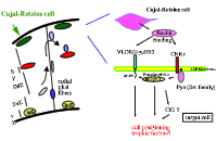
Cortical neurons are formed on the walls of cerebral ventricles and subsequently move towards the marginal zone (MZ) via radial glial fibers. After that, they take their final patterns of spatial arrangement under the influence of Reelin, which is secreted from the Cajal-Retzius cells in MZ. The figure represents a Reelin-signaling cascade.
|
Senzaki, K., Ogawa, M., Yagi, T.
Proteins of the CNR Family Are Multiple Receptors for Reelin
Cell 99, pp. 635-647 (1999)
|
|
 |
 |
|
|