Keeping mouse memories in order is all in the timing
Hippocampus oscillations organize neural code
May 31, 2016
Cross-posted from Neurographic
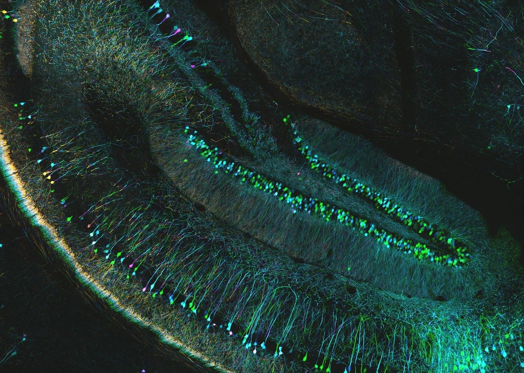
A stained section of mouse hippocampus seen through a confocal microscope.Image: ZEISS Microscopy, CC BY-NC-ND 2.0
“Well we know where we’re going, but we don’t know where we’ve been”
Mice from Tom McHugh’s lab can really relate to these lines from the Talking Heads’ song “Road to Nowhere”. The mental map of space in these mice is a little jumbled — each individual neuron is doing its job of signaling a location in the environment correctly, but putting them all together doesn’t give the mouse the right path from A to B.
It all comes down to the hippocampus, a part of the brain that is shaped a bit like its namesake, the seahorse. Sub-regions within the hippocampus rely on input from each other to update a representation of the environment as the mouse moves around. This input comes in the form of coordinated waves of brain cell activity; one of these wave patterns, called the theta cycle, runs eight times a second (8 Hz). Experiments in the McHugh lab found that the theta cycle, which is present in two regions called CA1 and CA3, is crucial for coordinating the ensemble of neurons.
The researchers genetically engineered mice to express tetanus toxin in the CA3 area of hippocampus. The neurons there were still active but this manipulation effectively silenced their communication to the downstream area of CA1, where the updating spatial map is located.
This illustration shows what’s going on in the brains of normal and mutant mice.
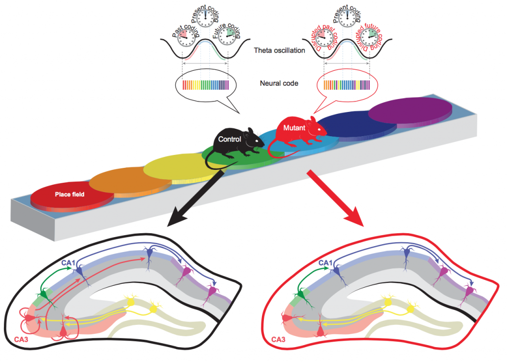
As control mice run along a track, the brain map of their environment is
regularly updated through the neural circuitry in the hippocampus (left side of image).
Without input from area CA3 in the hippocampus, the neural code that represents where
the mutant mouse has come from (past coding) and is going (future coding)
becomes disordered with only the current location remaining intact (right side of image).
Image: Steven Middleton/RIKEN Brain Science Institute
Mice in the experiment ran along a track. Where they’ve been, the current location, and where they’re going are represented by the rainbow of circles called ‘place fields’. These are the zones in the world represented by particular ‘place cells’ in the hippocampus, the discovery of which was recognized by a Nobel Prize in 2014. For the normal mouse on the left side of the image, the place fields line up in an orderly fashion one after the other in the neural code. This is achieved through the brain connections shown on the bottom left: neurons in CA3 mirror the waves of activity — the theta oscillations — that are sent to CA1 and help organize the neural code for the spatial map. Because of the experimental shutdown of CA3, the mutant mouse on the right has a disordered neural code. The neurons can tell the mouse its current location, but the information about the past and future locations are out of sequence.
The consequences for the mutant mice in this instance aren’t too severe: they aren’t navigating a maze but a one-way track, a literal ‘road to nowhere’. In a more complex environment, however, the temporal disorder of location memories could be problematic. Jumbled memories in conditions like Alzehimer’s disease may be tied to disrupted brain oscillations of the kind that were artificially generated in this experiment. Mouse or human, for knowing where you’re going and knowing where you’ve been, a correctly sequenced neural code is essential, but the greater contributions of rhythms in the brain to information organization still have to be worked out.
Middleton SJ, McHugh TJ (2016). Silencing CA3 disrupts temporal coding in the CA1 ensemble. Nature Neuroscience, DOI: 10.1038/nn.4311
Related Post
![]() RIKEN Press Release: The brain clock that keeps memories ticking
RIKEN Press Release: The brain clock that keeps memories ticking