  |
 |
 |
Columnar
structures in the human cerebral cortex
successfully observed from outside of the head
Cognitive Brain Mapping
Research Team |
|
 |
There
are high expectations for the elucidation of higher human brain functions, as
a result of accumulating experimental results from experimental animals and continuing
developments in noninvasive means of taking readings of neural activity. However,
the spatial precision of conventional non-invasive methods for recording readings
hase been on the order of 5 mm. With this degree of spatial precision, it has
been possible to determine where neural activity is raised to a higher position
with various mental activities, but impossible to confirm the mechanism by which
each of the positions in the brain achieves its respective functions. Our research
team in the Cognitive Brain Mapping Laboratory achieved spatial accuracy of 0.5
mm through improvements in magnetic resonance imaging technique, and was successful
in imaging the columnar cellular functional structure of the cerebral cortex.
The columns are small regions that extend in a direction perpendicular to the
surface of the cerebral cortex, and in which neurons with similar properties are
gathered. The breadth of the columns at the surface of the cerebral cortex is
approximately 0.5 mm in cats and monkeys. In the present study, the research team
imaged ocular dominance columns of the primary visual cortex, that is, the cells
gathered there that mainly receive input from the left eye, comprising the left
eye column, and the cells that mainly receive input from the right eye, comprising
the right eye column.
Using magnetic resonance imaging equipment, the team implemented a method that
determined heightened neuron activity by measuring increases in local blood flow
(functional magnetic resonance imaging method). Local blood flow increases reflexively
with increased neuron activity, and decreases levels of deoxidized hemoglobin
in capillaries. The decline in levels of deoxidized hemoglobin delays the attenuation
of proton magnetic resonance signals, thus increasing magnetic resonance signals
overall. The limitations on the spatial precision of functional magnetic resonance
imaging are ultimately determined by the space between capillaries (on the order
of 50 microns); however, due to the poor signal-to-noise ratio of the readings,
only highly imprecise results could in fact be obtained in the past. In order
to improve the signal-to-noise ratio, we developed various technologies -for example,
raising the static magnetic field to 4 Tesla, 2.5 times the level of conventional
technologies - and consequently increased spatial precision.
The human primary visual cortex extends along the sulcus known as the calcarine
sulcus, which stretches forward and back along the inner surface of the occipital
lobe of the cerebral hemisphere. The detailed shape of the brain varies among
individuals, but in most people the part of the cerebral cortex that extends along
the upper and lower walls of the calcarine sulcus is relatively smooth. Accordingly,
we adjusted the inclination and position of the imaging slice surfaces for individual
test subjects, so that the slices completely overlapped the cerebral cortex over
as wide a range as possible covering the upper or lower wall of the calcarine
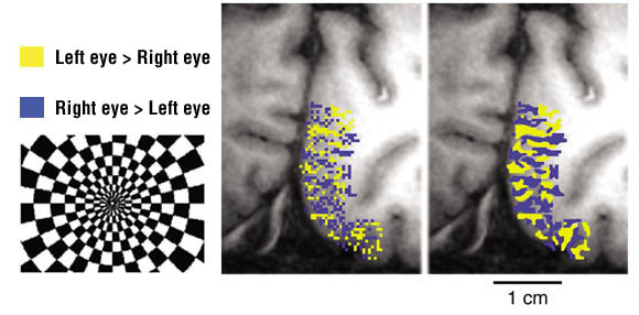
sulcus. For visual stimulation, we projected 8 black-white alternations per second,
in a black and white checkerboard pattern, onto the retina of one eye at a time
via an optic fiber bundle. A striped pattern was obtained by comparing the signal
distribution occurring in left eye stimuli with that occurring in right eye stimuli
(see Figure). The a striped pattern was similar to that of the ocular dominance
columns of monkeys, and the long axes of the bands were roughly perpendicular
to the boundary of the primary visual cortex (on the inner surface of the cerebral
hemisphere), as in monkeys. However, the average width of a human column was 1
mm, about twice the width of monkey columns.
In the present study, we noted that structures known to be present in monkeys
and cats are also present in humans. We plan to further improve the method, and
apply it to areas associated with the cerebral areas that are involved in uniquely
human higher brain functions. By identifying the stimuli and conditions that activate
each column, we will be able to infer information expression at the cellular level;
by comparing this with information expressed by adjacent columns, we will be able
to infer the details of information processing performed through interactions
between columns. This gives rise to the possibility of dramatically accelerating
research into the mechanisms of higher human brain functions. |
| |
 |
 |
Pattern of ocular dominance columns imaged by 4T fMRI in human primary visual
cortex
Neuron, Vol 32, 359-374, October 2001A aggregates in the presence or absence of zinc.
 |
|
 |
 |
|
|


