|
 |
 |
Elucidating
the mechanism by which organisms distinguish "belly" from "back".
-Discovery of a calcium signal target gene responsible for body axis-
Laboratory for Developmental
Neurobiology |
|
|
The
Developmental Neurobiology Team, working in cooperation with the Japan Science
and Technology Corporation (Kazuki Okimura, President) and the University of Tokyo,
has elucidated part of the calcium signal mechanism by which information that
determines "belly" and "back" in the early development of
organisms is transmitted. The discovery was the fruit of research by Takero Saneyoshi
and other researchers on the Developmental Neurobiology Team led by Katsuhiko
Mikoshiba.
The formation of an axis that distinguishes ventral and dorsal
plays an important role in processes such as neural tube development, by which
a single fertilized egg forms a cell population.The research group demonstrated
in the present study that the calcium signal acts as a ventralization signal by
acting on the calcium-dependent transcription regulator NF-AT, which is involved
in the immune system. They also found that NF-AT acts on the enzyme known as GSK-3 ,
which is related to the formation of back side dorsalization, and promotion of
ventralization. GSK-3 has also
been found to play a role in aging of the brain. ,
which is related to the formation of back side dorsalization, and promotion of
ventralization. GSK-3 has also
been found to play a role in aging of the brain.
These results demonstrate for the first time that the action of the calcium signal
in early development is deeply involved in basic biological phenomena such as
immune reactions and aging. These results, which were published in the British
science magazine Nature (May 16 issue), are expected to contribute greatly to
the acquisition of new information concerning morphological abnormalities, through
elucidation of the effects of substances such as drugs and endocrine disrupting
substances on the calcium signal, and are also expected to contribute to clinical
practice.
The Ca2+ signal has various ways of changing
the intracellular concentration of Ca2+, such
as transient increases, prolonged increases, and Ca2+
oscillation, in which the Ca2+ concentration
repeatedly rises and falls. It plays an important role in intracellular information
transmission by using the differences between these. On the other hand, it has
been known for some time that lithium, a drug for manic and depression patient,
induces dorsalization during the early development of organisms, but the mechanism
of lithium's dorsalization effect had long puzzled developmental biologists. The
Developmental Neurobiology Team has conducted research on the calcium signal and
inositol 1,4,5-trisphosphate (IP3), which is thought to have a particularly strong
relationship to the physiological activities of the brain, in the inositol metabolic
turnover pathway which is one of lithium's sites of action, and on its receptor
(IP3R), using a method of analysis based on experiments with model animals. As
a result of the study, in which we used an antibody that selectively inhibits
IP3R function, we found that when IP3R function is blocked on the future ventral
side, ventral cell fate switches to dorsal cell fate followed by the formation
of the secondary axis (the second back formed by switching the ventral side to
the dorsal side, in addition to the original dorsal side, is called the secondary
axis (Figure 1-1)).These results showed that the IP3-Ca2+
signal operates as the ventralization signal, and were published by Shoen Kume,
a researcher on the team, in the US journal Science (Kume et al., Science 278,
1940-1943 (1997)). The results prompted questions as to whether there is an IP3-Ca2+
gradient along the dorsoventral axis in early embryos, what molecules act upstream
and downstream from the IP3-Ca2+ signal transmission
system, and what other effects are related to, or interact with, ventralization
and dorsalization factors.
The nuclear factor of activated T-cells (NF-AT) is a transcription regulator that
decodes Ca2+ signal activity. It is controlled
by calcineurin (Cn), a Ca2+ calmodulin (CaM)-dependent
dephosphorylating enzyme. It has been found through analysis that the Cn/NF-AT
pathway is crucial to the activation of cells in the immune system. It is also
involved in hypercardia and skeletal muscle differentiation. Through the type
of expression that inhibits NF-AT transcription factor, the body axis (secondary
axis) is formed in the same way as when IP3 receptor is inhibited (Figure 1).
In addition, dorsalization (neural tube, etc.) is prevented by excess expression
(activation) of NF-AT (Figure 2). These results clearly showed that NF-AT transcription
regulator acts as a ventralization signal.
When activated NF-AT transcription regulator was co-expressed in an individual
whose secondary axis formed by inhibition of IP3 receptor, in order to investigate
the relationship between the IP3-Ca2+ signal
transmission system and Cn/NF-AT pathway, the secondary axis disappeared and the
individual shape was restored (Fig. 3). On the other hand, there is a pathway
which is considered to be completely independent, named the Wnt/ -catenin
pathway, which acts as the dorsalization signal, and in that pathway an enzyme,
glycogen synthase kinase 3
(GSK3-),
is active (Fig. 5). Inhibition of this GSK3-
causes switching of the ventral side to dorsal side (formation of the secondary
axis). This is because overexpression of GSK3-
restores formation of the secondary axis (Fig. 4, right panel), which occurs by
inhibition of a transcription factor, NF-AT (Fig. 4, Ieft panel). These results
indicate cross-talk between the ventralization signal of IP3 receptor and NF-AT
and dorsalization signal including GSK3-
(Fig. 5). -catenin
pathway, which acts as the dorsalization signal, and in that pathway an enzyme,
glycogen synthase kinase 3
(GSK3-),
is active (Fig. 5). Inhibition of this GSK3-
causes switching of the ventral side to dorsal side (formation of the secondary
axis). This is because overexpression of GSK3-
restores formation of the secondary axis (Fig. 4, right panel), which occurs by
inhibition of a transcription factor, NF-AT (Fig. 4, Ieft panel). These results
indicate cross-talk between the ventralization signal of IP3 receptor and NF-AT
and dorsalization signal including GSK3-
(Fig. 5).
GSK3-
is one of the enzymes that have been found to be active in neurons when neuron
death is induced through such causes as aging of the brain, and it has been shown
to be related to Alzheimer's disease. The results that we obtained in the present
study, indicating that abnormalities in body axis development are eliminated through
activity of this enzyme, also yielded the very interesting result that GSK3-
activity is intimately related to early development, in addition to aging. The
discovery of the fact that calcium messenger activity is closely related to the
formation of the body during early development may lead to new information clarifying
the mechanisms by which healthy brains and bodies develop. It should also contribute
greatly to the development of pharmaceuticals with fewer side effects, through
analysis of the effects of drugs and other substances on the calcium messenger,
and also to clinical practice, by making it possible to examine the effects of
endocrine disrupting substances on the calcium messenger. |
Nature 417, 295 - 299 (2002)
Takeo Saneyoshi, Shoen Kume, Yoshihara Amasaki, Katsuhiko Mikoshiba
|
|
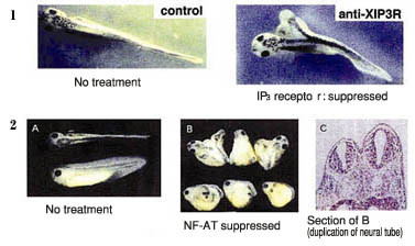 |
Figure 1: Change from ventral to dorsal.
1.A secondary axis is formed when IP3 receptor is suppressed. (Release of Ca2+
from the IP3 re ceptor determines the dorso-ventral axis.)
2.A secondary axis is formed when NF-AT transcription factor is suppressed (B
and C in the figure). |
|
|
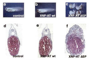 |
Figure 2: Dorsalization (neural tube, etc.) disappears and turns to ventralization
when NF-AT transcription factor is overactivated (c,f). |
|
|
 |
|
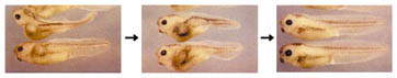
Figure 3: Secondary axis formation (left) through inhibition of IP3 receptor recovers
by activated NF-AT (right). |
|
|
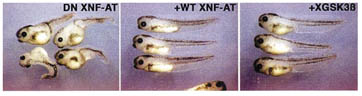 |
Figure 4: Secondary axis formation (left) due to suppression of NF-AT transcription
factor recovers by addition of NF-AT (middle) and further recovers due to GSK-3 overexpression (right).Note:3 Figures below are omitted.
 |
|
|
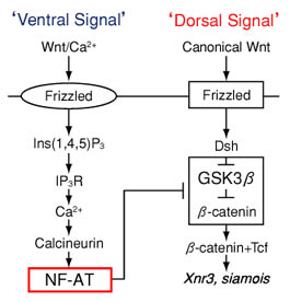 |
Figure 2: Dorsalization (neural tube, etc.) disappears and turns to ventralization
when NF-AT transcription factor is overactivated (c,f). |
|
 |
 |
|
|