|
 |
 |
Requirement
for Hippocampal CA3 NMDA
Receptors in Associative Memory Recall
RIKEN-MIT Neuroscience
Research Center
Gene manipulation in mouse and applications to the study of memory |
|
|
For
the first time, researchers at the RIKEN-MIT Neuroscience Research Center (RMNRC)
have identified a gene involved in the retrieval of long-term memories. The study
appeared in the May 30 online edition of Science. Nobel laureate Susumu Tonegawa,
director of the Picower Center for Learning and Memory at MIT and an RMNRC Investigator,
said his latest discovery may lead to drugs that would counter the memory loss
that plagues Alzheimer's victims and relieve middle-agers' "senior moments."
Tonegawa's laboratory explores the molecular, cellular and network mechanisms
underlying memory. He has pioneered the development of a genetic technology that
permits researchers to create a new strain of mice in which a specific gene is
knocked out in a specific area of the brain. In previous studies, Tonegawa and
colleagues at MIT used the genetically engineered mice to show that a gene for
the NMDA receptor in a region of the hippocampus called CA1 plays a crucial role
in the formation of a long-term memory. For these studies, Tonegawa generated
and analyzed mice with a genetically altered hippocampus, a brain structure that
has been shown to play a crucial role in common types of memories.
RETRIEVING MEMORIES
The formation of a long-term memory starts with input through one of the five
senses. We store an endless array of information as memories. Much of it is tucked
away in the brain's cellular networks at the subconscious level. To recall a memory,
we need cues to activate the memory network and bring the stored information to
the conscious level. "Very limited cues are sufficient to trigger a chain
reaction that permits us to become aware of the rich and detailed content of a
memory, "Tonegawa said. "This phenomenon is called pattern completion
because it reflects cellular processes accompanying memory retrieval in which
reactivation of a pattern of cellular connections harboring memory is completed
by very limited input."
The hippocampus is divided into three sections. This time, Tonegawa looked at
cells in the CA3 region, in which individual cells work in a so-called auto-association
network by sending signals back to each other as well as forwarding them to other
cells. Mathematical models suggest that through the modification of cellular connections,
memories stored in such a network can be efficiently accessed for the purpose
of pattern completion. Activity passed between cells through these connections
can replace information that might be missing in the input. In this way, the network
is able to "fill in" or retrieve complete memories given only a subset
of the original cues that were present when it was formed. "When people get
old, they complain they are losing their memory. In fact, in many cases they are
losing their ability to recall memories, "Tonegawa said. "It's the tip-of-the-tongue
phenomenon-if they had more triggers, they often can recall the facts. "The
same is true in the early phase of Alzheimer's disease. "It's not that they
lost the memory. It's that they have a retrieval problem,"he said.
MICE AND MEMORY CUES
In the experiment, mice were given a learning and memory challenge: locate a
submerged platform hidden from view in a pool of water. Although mice can swim,
they don't like water, so they immediately searched for a way out. After swimming
around randomly, they would find the platform and climb out. Visual cues placed
around the tank helped the mice orient themselves.
Each mouse that repeated the task several times a day for a week got better at
finding the platform, until they got to a point where they swam straight to the
platform. After the animal had learned the task, several of the visual cues outside
the tank were removed. Even with only one visual cue remaining, normal mice remembered
the location of the platform quickly and easily.But with only one cue, mice with
altered NMDA receptors in the CA3 area of the hippocampus could not find the platform.
They could only find it with all four cues intact.
PLACE CELL DEFICIT
Tonegawa says this study is striking because it provides evidence at the behavioral
level as well as the molecular level. To provide insight into the link between
these levels, Wilson has devised a method to monitor the actions of dozens of
individual neurons in real time. With electrodes that reach into the depths of
the hippocampus, the researchers can actually see memories being formed in the
brains of their mice subjects. Each memory creates a unique pattern of activity
in the hippocampus. Individual cells-called place cells-fire in response to memory
activation of different places.
Using these recording techniques and in a collaboration with another professor
at RMNRC Dr. Matthew Wilson, postdoctoral fellow in Dr. Tonegawa's laboratory,
Dr. Kazu Nakazawa, was the researchers were able to identify the characteristics
of neural activity corresponding to the partially cued reactivation of previously
formed memories, or pattern completion. When animals explored an environment in
which all four visual cues were present, the patterns of place cell activity in
the genetically altered mice appeared to be normal. But when cues were removed,
the cells responded much more weakly than cells in normal animals. The weakened
response was attributed to the inability of the CA3 area to successfully retrieve
the complete memory of that environment given only partial cues. This result provides
a direct measurement of how failed memory retrieval might appear at the level
of individual neurons, and therefore provide the basis for improving memory under
these conditions.
A CRUCIAL PATHWAY
The density of NMDA receptors is reduced in older people. The CA3 region of the
hippocampus is known to be most vulnerable to plaque formation in the brains of
Alzheimer's patients. "By understanding how memory retrieval works at the
molecular level, this could be the basis for drug development in the future,"
Tonegawa said. "It's important not only for Alzheimer's patients, but for
prolonging the ability of aging people to recall memories and learned facts." |
|
|
 |
|
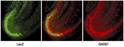
Fig1. Immunohistological Staining of the Hippocampal
Slices from the Mutant Mice: The green color represents CA3 cells in which the
gene knockout has occurred. The red color on the right represents inhibitory interneurons
in CA3. The merged picture in the middle in which the area stained green is largely
separated from the area stained red indicates that the knockout is restricted
to excitatory pyramidal cells in CA3. |
|
|
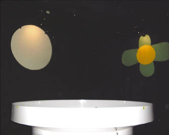 |
Fig2. The Morris Watermaze
Setup: The swimming pool (white at the bottom) filled with opaque water was set
in a dim lighted room surrounded by a black curtain on which four (only two are
shown) brightly lighted patterns were hung at four slides as the distal visual
cues. In the pattern completion experiment three out of the four cues were removed
during memory testing sessions. |
Science,
Vol. 297, Issue 5579, 211-218, July 12, 2002
Kazu Nakazawa, Michael C. Quirk, Raymond A. Chitwood, Masahiko Watanabe, Mark
F. Yeckel, Linus D. Sun, Akira Kato, Candice A. Carr, Daniel Johnston, Matthew
A. Wilson, Susumu Tonegawa |
|
 |
 |
|
|