|
 |
 |
How
are object images represented in our brains?
Laboratory for Integrative
Neural Systems |
|
|
How
do our brains perceive complex objects? Using macaque monkeys, The Laboratory
for Integrative Neural Systems is striving to explain this mechanism.
Macaque monkeys are frequently used to model humans in vision research, since
they have similar visual systems to ours. In the temporal lobe visual association
area of macaque monkeys, there are clusters of neurons, comprising "feature
columns", that respond to specific features of fairly complex objects. Using
optical imaging (a technology for using light to capture neural activity), The
Laboratory for Integrative Neural Systems was able to observe these columns as
spots scattered on the surface of the brain.
When a monkey was shown an image of a complex object, many spots (active columns)
appeared on the surface of the brain, but when the object was disassembled and
the simplified features were shown to the monkey, some of the spots disappeared
(Figure 1). Upon investigation of the properties of neurons in individual spots,
we found that each spot is related to specific features that are included in the
image of an object (Figure 2). We surmised that the brain disassembles complex
objects and expresses the images using feature column combinations that correspond
to respective partial features.
A very interesting finding is that individual features of object images are explicitly
expressed when spots are activated and may also be implicitly expressed through
the non-activation of spots. For example, the fire extinguisher in Figure 2 is
a rotund figure next to the hose, which appears to be round. In the temporal lobe
visual association area, this rotundity is expressed through the non-activation
of certain spots (blue spots, in Figure 2). This process, the combination of active
and dormant columns, for image expression is more complex than a simple combination
of active columns as was previously assumed. |
Tsunoda K, Yamane Y, Nishizaki M, and Tanifuji M: Complex objects are represented
in macaque inferotemporal cortex by the combination of feature columns. Nature
Neuroscience 4, 832-838, 2001
| |
|
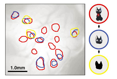 |
Figure
1: Arrangement of spots appearing on the surface of the brain when the features
lined up on the right were shown. The red, blue, and yellow spots indicate spots
that appeared in response to the entire cat image, the head only, and its silhouette
only, respectively. As the features were made simpler, spots that had appeared,
vanished. |
| |
|
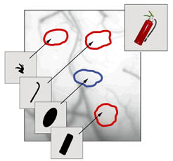 |
Figure
2: There were 4 spots appearing within the area measured, that were associated
with the fire extinguisher in the upper right. Among these, the red spots were
activated in response to partial features of the fire extinguisher. The blue spots
indicate response to rectangular images, but not to more oval shapes. These spots
were not activated in response to the fire extinguisher in the upper right; thereby,
they impart an overall sense of rotundity to the object image (a fire extinguisher).
It appears that object images are formed from the combination of columns that
are and columns that are not activated. |
|
 |
 |
|
|