


Thin layers of cells along the inside of the eye form the retina which is crucial for vision. The diseases in retina often cause many of the problems affecting normal sight; therefore, mapping retinal functions will lead to early discovery and the precise diagnosis of retinal diseases. Our laboratory, in cooperation with Laboratory of Visual Physiology, National Institute of Sensory Organs in Tokyo, observed the distribution of neural activity in a monkey's retina using a novel non-invasive imaging technique.
Background
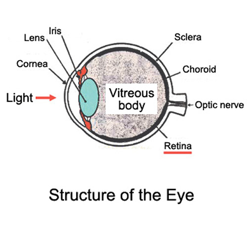
The retina is the membrane-like nerve tissue of the fundus in the human eye, that works much like film in still cameras (Fig. 1). They both absorb light and convert that light into electrical signals. Reduced function of photoreceptor cells in the retina leads to blindness or a reduced visual field. Traditional optical examinations rely on report of the subjects about their eyesight and the range of the visual field. Alternatively, electrophysiological methods that capture electric signals from the neurons in the retina are used, but these methods do not provide enough spatial resolution to diagnose retinal diseases or require long time to do so. We successfully created a detailed functional topography (neural activity distribution chart) of photoreceptor cells in primates using common optical imaging technology that enable us to objectively visualize the function of photoreceptor cells in a non-invasive way.
Research Results
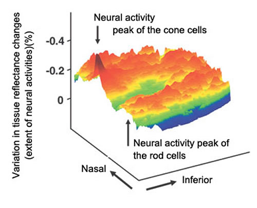
Optical imaging captures small changes in tissue reflectance caused by neural activity, and could be capable of mapping neural activity in human retina. We examined this possibility by using a monkey as a model animal, since retinal structure and function of rhesus monkeys is nearly identical to humans. We attached a video camera to a fundus camera designed for humans and observed changes of reflectance in retina with invisible infra-red light (having a wavelength of 800nm or more). The flash stimulus with visible light elicited neural activity in retina of an anesthetized monkey that was followed by reflectance change of retina. Changes in reflectance before and after stimulation were used to calculate the topography of that neural activity.
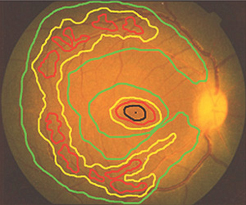
Our topographical results had a high peak corresponding to the responses of the cone photoreceptor cells at the center of the retina and a ring-shaped peak that corresponded to the responses of the rod photoreceptor cells in the peripheral area (Fig. 2). The findings were consistent with anatomical and psychophysical data of cone and rod distribution in human retina. We also found that the intrinsic signal was reduced in the area of artificial lesion.
This simple, novel technique for topographical analysis, which we are calling functional retinography, provides a non-invasive method for imaging the functional topography of the cones and rods. It is a quick way to measure the intrinsic signals of the retina.
Future Directions
Vision declines with age, and in more rapidly aging societies the number of people with vision-associated problems will increase significantly. Developing clinical applications of functional retinography is already underway. Once it can be put into practical use, we hope that early discovery of diseases, including macular degeneration and retinopathy of prematurity, that threaten eyesight will be possible. We also expect better evaluation of treatment effectiveness through this technology.
