


Functional asymmetry in cerebral hemispheres of the human brain is a characteristic of a variety of cognitive tasks, including the processing of speech and perception. Recent studies have also found asymmetric activation in response to external stimuli in the sub-cortical structures such as limbic system. Upon seeing a fearful face, for example, one side of the amygdala will show activation. These asymmetries are not unique to humans and may be a universal feature of nervous systems across species. Chickens, fish, toads show preferential use of one visual field for specific tasks, a preference that might extend to all vertebrates. Increasing evidence suggests that lateralization in information processing in the brain is evolutionarily conserved.
Surprisingly, the neuronal connectivity patterns that regulate this functional asymmetry remain poorly understood. How, for instance, is specific information conveyed from the left or right sensory processing structures to the motor regions of the brain. As motor output triggered by lateralized brain activity involves both sides of the body, circuitry that conveys that information from left or right neural structures to nuclei on both sides of the brain should exist. Nevertheless, the location of such circuits structures, if they exist, had yet to be identified.
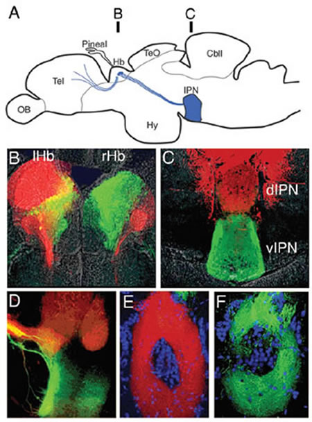
We uncovered a novel characteristic of neural circuitry that might provide a mechanism that shuttles information from the left and right sides of the brain to specific dorso-ventral (DV) regions of a bilaterally positioned target nucleus by analyzing the output circuitry of the lateralized habenular nuclei in zebrafish. The habenulae, evolutionarily highly conserved parts of a conduction pathway within the limbic system, connect telencephalic nuclei to the interpeduncular nucleus (IPN) of the midbrain. These nuclei probably regulate dopaminergic and serotonergic neuronal activities. Using zebrafish, we demonstrated asymmetric connectivity in the habenulo-interpeduncular projection: a stereotypic, topographic projection from left-sided habenular axons predominantly to the dorsal region of the IPN and from right-sided habenular axons predominantly to the ventral IPN (Fig. 1). A prominent left-right (LR) difference in the size ratio of the medial and lateral sub-nuclei of the habenulae can account for this asymmetry because each sub-nuclei projects to either a specific ventral or dorsal IPN target (Fig. 1).
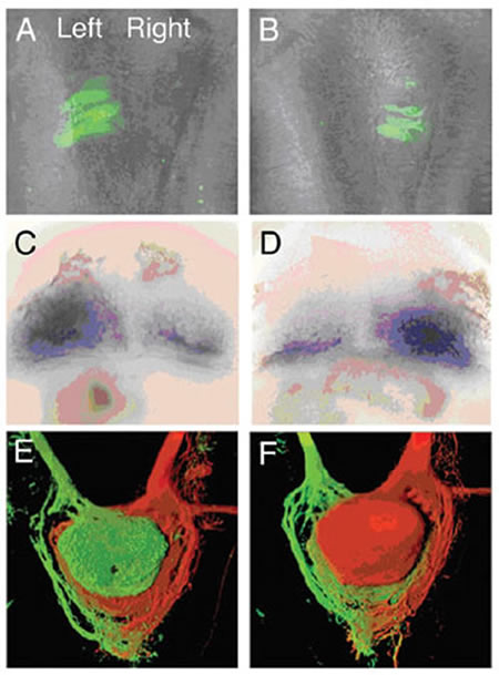
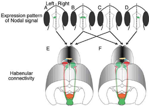
The Nodal signaling pathway regulates laterality decisions in the viscera. In zebrafish, unilateral activation of this pathway in the left brain specifies lateral asymmetry in habenular size (Fig. 2). Asymmetric Nodal signaling also determines the DV polarity of innervations of the IPN by left and right habenular axons (Fig. 2), however axon terminal segregation can still occur in mutant IPN if Nodal signaling is symmetric or absent (Fig. 3). The latter finding is consistent with Nodal pathway activity for coordinated lateralization in the circuitry between all individuals at the population level.
In this way, we successfully identified a mechanism by which information that is distributed between the left and right sides of the brain is transmitted bilaterally without losing LR coding. We have also started to elucidate the signaling pathways that direct the establishment of lateralized circuitry.
