


Our laboratory successfully imaged senile plaque, a pathological hallmark of Alzheimer's disease, in living mouse brains using a fluorine compound and magnetic resonance imaging (MRI). This technique will help efforts to develop methods for early diagnoses of Alzheimer's disease as well as to expose those mechanisms linked to disease onset and to assess suitable therapies that target senile plaques.
Background
Alzheimer's disease is one of the leading causes of dementia, yet it is currently impossible to accurately diagnose Alzheimer's disease in patients while they are alive. Moreover, radical treatments remain elusive. Pathological analyses of the brains of patients with Alzheimer's disease identified senile plaques and neurofibrillary tangles as the key characteristics of the disease. Senile plaques are extracellular aggregates of amyloid-β peptide (Aβ) while tangles are deposits of pathological tau protein found inside neurons. In Alzheimer's disease, Aβ self-aggregates into fibers by forming a secondary structure called a β-sheet. The extracellular accumulation of amyloid proteins can begin in the brain 20 to 30 years before clinical symptoms appear. Therefore, it may be possible to diagnose Alzheimer's disease well before those symptoms emerge by non-invasively detecting amyloid accumulation in living brains. As research has shown that amyloid in the brain also contributes to neurodegeneration, targeting ways to suppress amyloid-induced neuronal insults would be novel therapeutic approaches to these neurodegenerative diseases as well. In vivo detection of cerebral amyloid may also allow the effects of those therapeutics candidates to be observed and analyzed over time.
Recent Achievements
While it has been possible to detect senile plaques in vivo using radioactive chemicals and positron emission tomography, costly radiochemical synthesis and strict regulation for radioactive materials may hamper popularization of such approaches. So our laboratory focused on a detection technique using MRI. Most MRI approaches exploit the magnetic resonance of hydrogen nuclei (protons), but fluorine atoms, among others, can also be used as chemical tags in the body and imaged with a fluorine MRI scan that then quantifies the internal distribution of fluorine. Unlike hydrogen, fluorine does not naturally exist in living organisms, so any signal obtained in the scan would be associated with the fluorine-tagged compound.
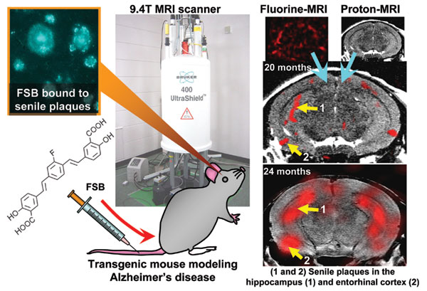
Working together with Dojindo Laboratories, we created a fluorine-containing compound with an affinity for amyloid that we called FSB* and attempted a fluorine MRI scan. As FSB is a fluorescent compound, we first stained slices from the brains of Alzheimer's disease patients and mouse models with Aβ plaque accumulation. We identified senile plaques with the fluorescent stain and confirmed that FSB binds specifically to amyloid. Next, we injected FSB into the veins of mice and examined dissected brain samples under a microscope. FSB was transferred to the brain and labeled senile plaques in these animals (see Figure). The blood brain barrier prevents most chemicals from penetrating the brain; however, FSB has a relatively low molecular weight and high lipophilicity allowing it to pass this protective barrier. Finally, we used the fluorine MRI to scan FSB-treated mice. A fluorine signal was detected in regions corresponding to typical senile plaque build-up, namely in the hippocampus and entorhinal cortex (see Figure). The MRI scanner had a 9.4-Tesla magnetic field that was designed specifically to image the mouse brain with high spatial resolution and can capture small structures between 50 and 100μm (see Figure). Knowing that senile plaque accumulates with age, we tested to see if the fluorine signal also increases accordingly. It did, and so we concluded that use of FSB for fluorine MRI scans can provide reliable quantitative assessments of senile plaque development in the brain.
Future Expectations
The success of our research in detecting amyloid accumulation in brain of a mouse model of Alzheimer's disease using MRI should provide a convenient way to evaluate the efficacy of various therapies in mice. Fluorine MRI can also be used to observe the progression of neurodegeneration and, one day, to identify when amyloid deposition occurs within that progression. This would eventually reveal the molecular mechanisms mediating the onset of Alzheimer's disease.
However, sensitivity of fluorine MRI is still not high enough to detect the distribution of trace amounts of chemicals. We had to administer high dosages of FSB (20mg/kg) before we could conduct a reliable fluorine MRI scan. Moreover, applications of the present method to human subjects poses significant challenges, However, we believe that clinical diagnoses in the future with this method will be enabled by further technical innovations, including development of an amyloid-specific compound with a higher fluorine atom content and more efficient transfer to the brain, as well as improvements of MR detection coil.
* : (trans,trans)-1-fluoro-2,5-bis-(3-hydroxycarbonyl-4-hydroxy) styrylbenzene.
