


Introduction
The human brain has about 14 billion neurons and over ten times that number of glia cells that exist within a highly complex structure that performs multiple functions. From the moment it is fertilized, an egg repeatedly divides and differentiates over the course of gestation. Eventually, the brain forms. That process of brain formation is our focus, through which we hope to understand the brain.
Sonic Hedgehog (Shh) Protein in Brain Development
A fertilized egg will repeatedly divided to become first an embryo, then a fetus. Part of the embryo forms a tube, called the neural tube, that is the start of the central nervous system: brain and spinal cord. The developmental from the neural tube to the brain may help explain the brain. We are investigating the mechanisms that determine the midline structure of the brain, the first crucial step in development. We are also trying to understand how neural stem cells emerge and develop.
A secreted protein called sonic hedgehog (Shh) currently commands our attention. This protein, which is essential for healthy brain development, maintains brain functions after birth. Constructing tissue with a large number of cells requires coordination and role allocation among the various groups of cells. For brain development, it is becoming clear that cell-to-cell interactions are supported by many molecular events are essential to that process. Shh may participate in those intercellular interactions. Efforts by our laboratory and others have shown Shh's roles in determining the midline structure and in directing the development of neural stem cells. We would like to reveal the functions and roles of other molecules involved in Shh protein signaling cascade that leads to the proper development of brain tissue.
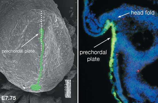
An SEM micrograph of the frontal view of a mouse embryo (fetal age 7.75 days) Left: Shh protein is green. Right: Enlarged, cross-section of an embryo at the same developmental day. The dotted line in the micrograph (left) shows the region: Shh antibody reveals Shh. The part that will become the brain (head fold) is followed by the perchordal plate. Shh (in green) that is expressed in the prechordal plate induces midline structure formation.

Sorting at the Mid-Line
The mid-line structure divides the brain in half: to produce the left and right hemispheres. The human embryo's midline structural development occurs about 3 weeks after the fertilization. Before this event, the future brain is single, undifferentiated layer of cells that is called the neural plate. Underneath these cells is by the entodermal cell layer. A group of cells located immediately below the center of the neural plate secretes Shh protein (Fig.1). The formation of the midline structure begins with this secretion. Shh protein secretion triggers Shh expression in cells in the neural plate. This secretion ultimately determines the balance between excitatory and inhibitory neurons as the brain continues to form.
Anomalies in Midline Sructure Development in Humans
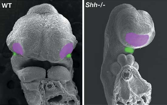
Holoprosencephaly is a congenital developmental anomaly in which the midline structure develops poorly or not at all. An incomplete midline structure and cyclopia can result when hemispheres are not formed (Fig.2). Either genetic or environmental factors can cause this developmental disorder. A mutation in the Shh gene was implicated in 1996. Drinking alcohol during pregnancy or being diabetic can also increase the risk that the embryo may develop holoprosencephaly. These environmental risk factors, we discovered, prevent the Shh signaling cascade, thereby impairing development of the midline. Hence, a possible explanation for environmentally induced holoprosencephaly is the failure of the hedgehog signal.
Mechanism of Neural Stem Cell Development
Neurons and glia cells emerge from neural stem cells. These stem cells produce neurons at an early stage of development; glia cells come later. The mechanism regulating the timing of that staged production is still unknown. We are attempting to clarify the essential role of the Shh protein-signaling cascade in the development of normal neural stem cells.
Brain Tumor due to Abnormal Sonic Hedgehog Signal Transmission
Reports suggest that some cancers, including brain tumors, in humans and mice may be due to an anomaly in the SHH signaling cascade (Fig.3). Those molecules involved in the Shh signaling cascade can be seen in various cells of postnatal brains, including in the neural stem cells. Although cell division is inhibited after birth, cells may become cancerous when there is an anomaly in the Shh signaling cascade. We are planning expose how Shh failure might lead to cancer by studying mutant mice. We showed that environmental factors could interfere with Shh function. Together with the possible role of neural stem cells in maintaining normal brain function and Shh maintenance of those stem cells, we think that environmental factors may affect the Shh signal cascade. We would like to learn how environmental stimuli affects the SHH signal and how that would affect brain functions.
Conclusion
Our goal is to understand the mechanisms of brain development. We identified the developmental mechanism in this process that are permanently affected by environmental factors (including pre- and post-natal environments). These effects vary according to the environment. Our research will help explain how variations in brain development mechanisms, i.e. environmental factors, arise and identify environmental risks in development of the brain in maternal and postnatal periods.
