


Large networks of interconnected neurons enable learning, memory, and motor control in the brain. A basic neuron has a soma (cell body), an axon, and dendrites. To make connections with other neurons, a neuron sends and receives neurotransmitters like glutamic acid through its axon and dendrites. Calcium serves as an important intracellular messenger in these exchanges. However, to work effectively, the levels of calcium concentration must be carefully regulated over time. Too much calcium, for example, can lead to cell death. Inositol thriphosphate (IP3) receptors are intracellular ion channels that regulate intracellular calcium concentration by removing calcium from the intercellular stores in response to extracellular signals. There are three kinds of IP3 receptor and the type 1 IP3 receptor (IP3R1) is expressed in large quantities in brain neurons.

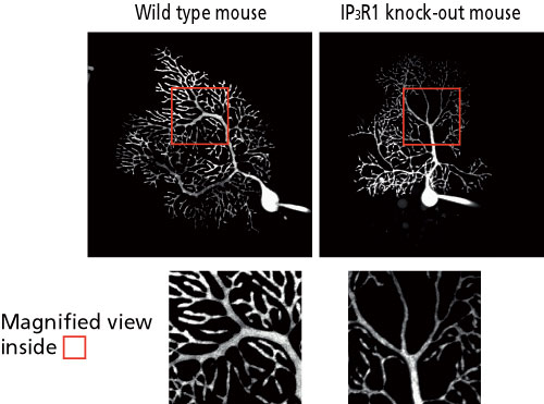
In our laboratory, we cultured cells taken from the cerebellums from wild-type and IP3R1 knockout mice. We compared Purkinje cells arborization in these areas and found that the he IP3R1 knockout mice showed less branching and longer dendrites than the wild-type mice (Fig. 1).
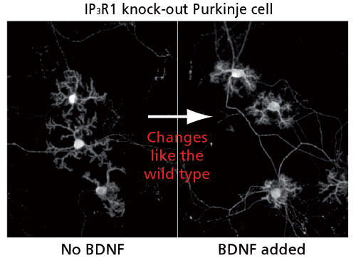
Next, we look at the possible contribution of cerebellar granule cells, a major neuron that transmit messages to the Purkinje cells, in controlling dendritic outgrowth in Purkinje cells. First, we showed that, contrary to previous assumptions, IP3R1 is most abundantly expressed in granule cells. Granule cells produce Brain-Derived Neurotrophic Factor (BDNF), which affects dendritic arborization in response to external signals. Since the external stimuli may also promote calcium mobilization via IP3R1, we studied the relationship between calcium mobilization and BDNF expression. Calcium mobilization in response to exogenous signals was rarely observed in granule cells of the IP3R1 knockout mice and that increases in BDNF expression were low. And when we added BDNF to the cerebellar cells of IP3R1 knockout mice, the dendrite outgrowth returned to normal levels (Fig. 2).
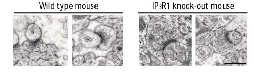
We injected Purkinje cell dendrites with a fluorescent dye to observe dendrite arborization under a microscope. The Purkinje cells of the wild-type mice presented an orderly branching while those of the IP3R1 knockout mice were abnormal (Fig. 3). This decreased branching of the dendrites supports our findings from the in vitro studies. Our electron microscope observations also indicate abnormal vesicle accumulations at the extremities of the axons of the cerebellar granule cells of the IP3R1 knockout mice. These accumulations appear to be neurotransmitter and, if this were the case, it would suggest that communication between the granule cells and the Purkinje cells (Fig. 4) is impaired in these knockout mice.
When these findings are taken together with the earlier finding about BDNF, we can conclude that BDNF produced by the IP3R1 receptors in the granule cells probably controls neurotransmitter release by acting on the axons of the granule cells themselves.
In the future, we want to create mice lacking IP3 Receptors in a neuron specific manner as well as mice lacking all three IP3 receptors so we will be able to completely understand all the functions of IP3 receptors in neurons. We will also be able to see these molecules contribute to neural network formation and thereby enable more complex brain functions.
