The contributions of fMRI (functional Magnetic Resonance Imaging) in exposing the relationship between neuronal diversity across brain regions and brain function are invaluable. However, we still lack a way to understand how EEG (Electroencephalography) measurements of electrical activity in neurons may correspond with data captured using fMRI. If explored, this situation could lead to the development of new experimental techniques. In our laboratory, we study various functions of the brain by exploring the role that synchronization plays in establishing a flexible, functional neural network. Revealing the dynamic properties of a neural circuit is one of our key interests. By measuring brain activity with fMRI and a time-domain resolution of scalp-recorded brain waves with EEG simultaneously, we can get information about that activity with high spatial accuracy.
Scalp-recorded brain waves between 4 to 8 Hertz are called theta-rhythms. In 1971, Ishihara et al. found theta rhythms in the frontal-parietal regions increase as a person attempts to solve a mathematical or a geometrical problem quickly and accurately. These waves, more precisely called frontal-midline theta (fm theta), probably emanate from in or around the anterior cingulate cortex. fMRI data from humans and EEG experiments with animals show that activity in this area is not triggered by any specific action. Rather, the area seems to monitor of the execution status of various actions, or, out another way, this region has centralized executive function. From the viewpoint of our vibration synchronization hypothesis, if fm theta activity participates in the monitoring function, we should observe synchronization of theta waves between the monitoring site, and those it is monitoring.
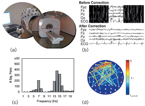
To verify this theory, we measured the theta waves observed in the fm scalp to identify the kind of circuit formation they are engaged in, as well as the activities of the entire brain. We conducted a simultaneous EEG/fMRI measurement experiment using mental arithmetic calculations, which are known to generate fm theta waves.
Hiroaki Mizuhara (presently at Kyoto University) led the project in cooperation with Tokyo Denki University. To analyze the results, a baseline was set by indexing a certain amount of brain waves and then calculating the slow variable "expected BOLD" (that is the value expected from the time series) and accessing the correlations between the expected BOLD value and the BOLD values measured at various brain sites.
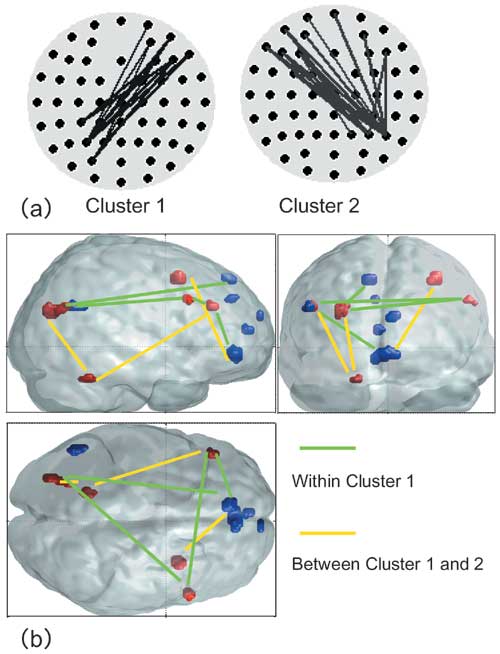
First, we extracted the time series of phases having the same frequency components from a pair of arbitrarily selected electrodes to obtain the degree of the phase difference between the two electrodes. These would be the phase synchronization variables and we could identify the electrode pairs where variables increased dependently (NeuroImage 2005, 2007). We identified several electrode pairs with the same phase synchronization at 7 Hz (theta rhythm) at remote sites and categorized these electrode pairs into clusters. Phase synchronization for each cluster was set as an index of brain waves and applied it to the analysis of correlation between fMRI and BOLD (NeuroImage 2007).
The results revealed the neural circuitry between the sites from which the fm theta emerged and showed how several domains are connected and disconnected through theta phase synchronization. With a successive subtraction process, using the working memory of the above experiment, the sites belonging to the special working memory and those associated with the motions involving the cerebellum appeared alternately. In addition, further analysis showed that the circuitry between the cortical layer and the striatum appears following the phase synchronization at 14 Hz (beta rhythms) during mental arithmetic calculation. The above results show an aspect of brain functions that is based on mutual monitoring of activity by means of synchronizations in multiple frequency bands.
2)Mizuhara H., et al: NeuroImage Vol. 27 No. 3, pp. 553-563 (2005)
3)Mizuhara H., Yamaguchi Y.: NeuroImage Vol. 36 No. 1, pp. 232-244 (2007)