


Background
Living cell membranes are composed of lipids and proteins, but these molecules are not uniformly distributed certain molecules are assembled or scattered. The clustering of molecules with specific functions leads to localized functional differentiation of the cell membrane, thereby enabling region-specific transmission of biological signals. The cell membrane includes lipid microdomains, which are composed mainly of cholesterol and sphingolipids, and are currently attracting much interest among researchers. Lipid microdomains, which vary in size from tens to hundreds of nanometers in diameter, repeatedly scatter and reassemble every second, and a substantial number of essential signal-transducing molecules gather there. Because of this, lipid microdomains are considered to serve as platforms for intracellular signal transduction. They are also involved in many biological phenomena, such as immune system function and Alzheimer's disease.
The growth cone, a tip of an elongating neurite, expresses cell adhesion molecules. These adhesion molecules on the growth cone react to their surroundings and generate intracellular signals that regulate neurite elongation. As mentioned above, lipid microdomains are also present in the cell membrane of growth cones. However, it has been difficult to examine whether or not lipid microdomains are involved in neurite elongation, partly due to lack of a functional inactivation technique that would be specific for lipid microdomains in neurites. Thus, the development of a technique enabling rapid intervention in the functions of lipid microdomains at particular locations has long been awaited.
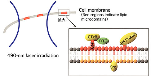
Lipid microdomains consist of specific lipids and proteins (for example, GPI proteins), and are enriched in signaling components (for example, Src). Therefore, they are regarded as platforms for intracellular signal transduction. FITC-CTxB specifically recognizes and binds to GM1 gangliosides expressed within lipid microdomains. Upon laser irradiation, the FITC generates free radicals that destroy molecules in its close vicinity. In this way, lipid microdomains can be inactivated in a local subcellular area (laser-irradiated area).
Copyright Rockefeller University Press
Methods and Techniques
Yoko Nakai, researcher, and Hiroyuki Kamiguchi, team leader, Laboratory for Neuronal Growth Mechanisms attempted to disturb the functions of lipid micro-domains at particular locations, using micro-scale chromophore-assisted laser inactivation (micro-CALI), a technique by which the functions of specific molecules in irradiated areas are disturbed by means of microscope laser irradiation (Fig. 1). We prepared FITC-conjugated CTxB obtained by conjugating fluorescein isothiocyanate (FITC) to cholera toxin B subunits (CTxB), which specifically recognize glycosphingolipids (GM1) existing only in lipid microdomains. Then, by adding FITC-conjugated CTxB to cultured nerve cells, lipid microdomains of nerve cells were labeled with FITC, which generates free radicals upon laser irradiation at the wave length of around 490 nm. Taking advantage of this property of FITC, we could then destroy molecules within the close vicinity of FITC with free radicals by applying 490 nm laser irradiation to a particular area of the cell membrane. The spatial range of these free radicals is less than 5 nm, so the effect is confined to the lipid microdomains. We were able to demonstrate that laser irradiation for 30 seconds causes lipid microdomains to be inactivated
.
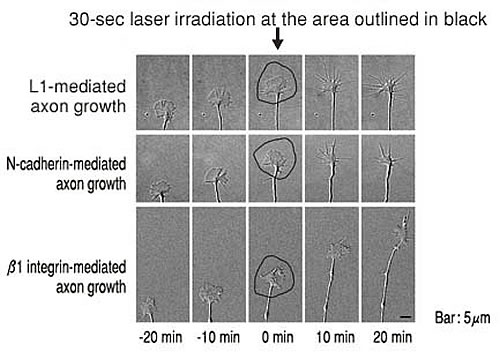
Next, we examined the involvement of lipid microdomains in neurite elongation. First, we analyzed the localization of adhesion molecules (L1, N-cadherin, 1 integrin) on the cell membrane. This revealed that L1 and N-cadherin are present both inside and outside lipid micro-domains, but 1 integrin exists only outside lipid micro-domains. Inactivation of lipid microdomains in the peripheral domain of the (growth cone) using micro-CALI resulted in the arrest of neurite elongation stimulated by L1 and N-cadherin but not by 1 integrin (Fig. 2). Further analysis demonstrated that lipid microdomains in the peripheral domain of growth cones are essential for neurite elongation, but those in the central part of growth cones are not.
Future Prospect
We have succeeded in developing a new technique for rapid and location-selective intervention in the functions of lipid microdomains, thereby demonstrating the importance and functional localization of lipid microdomains involved in the process of neurite growth. This technique will provide essential data for the progress of cell biology, as lipid microdomains are known to be involved in many biological phenomena. We hope that the spread of this technique will contribute to breakthroughs in our understanding of region-specific functions within cells and tissues, and to the elucidation of localized functional differentiation and intracellular signal-transducing mechanisms of the cell membrane.
