


Researchers at the RIKEN-MIT Neuroscience Research Center are one step closer to understanding how brain synapses make chameleon-like changes in their structure and composition depending on the input they receive.
Yasunori Hayashi, Unit Leader of RIKEN-MIT Neuroscience Research Center, seeks to understand how brain cells accomplish their remarkable plasticity. His work on the shape-shifting cell protein called actin appeared in Nature Neuroscience. This knowledge may one day make it possible to enhance learning and memory by manipulating neurons at a molecular level.
In the fraction of a second one brain cell to communicate with another, a lot happens. Chemical neurotransmitters are released from the signaling side of the synapse and bind to the receiving side, which triggers certain proteins to be assembled or disassembled. Long-term changes in the structure of brain cells creates long-term memories and lifetime learning, while other changes destroy unneeded connections to eliminate unneeded information.
A cellular protein called actin is responsible for helping synapses keep their shape. Hayashi and colleagues speculate that actin works with other mechanisms to help synapses assemble proteins on the postsynaptic, receiving end of a transmission.
Actin itself is transformed from ball-like globular to stringy filament forms (G-actin and F-actin), which do radically different things during key brain processes. How actin behaves and what it does is not well understood because no one has been able to see this conversion in synapses in living neurons.
Using a method called fluorescent resonance energy transfer (FRET) in combination with a state-of-art microscopic technique called two-photon laser scanning microscope, Hayashi was able for the first time to observe the change in equilibrium between G-actin and F-actin. By attaching a protein derived from jellyfish, he modified actin so that G-actin glows blue and F-actin in yellow. Change in color from blue to yellow means a conversion from G-actin to F-actin. “Without FRET, we couldn t see this kind of change,” Hayashi said.
Hayashi s laboratory zapped the fibers of a pre-synaptic cell in the rat hippocampus, the brain region associated with formation of new memory, with intense electrical stimulation. This initiates a biochemical process creating a physical change that can last for hours or even days in the post-synaptic receiving cell, either potentiation or depotentiation. These two key brain processes are long-term potentiation (LTP) and long-term depression (LTD), which many believe are the basis for learning and memory. LTP causes long-lasting changes by helping build new connections among brain cells and LTD helps destroy unneeded connections. The low-frequency stimulation created a reaction like LTD, while the intense electrical stimulation mimicked LTP.
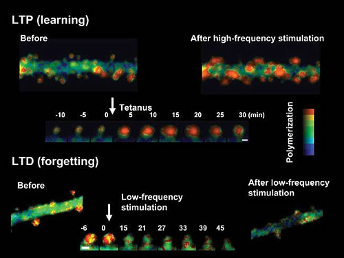
Hayashi and colleagues Ken-Ichi Okamoto, postdoctoral associate in the Picower Center for Learning and Memory, and Takeharu Nagai and Atsushi Miyawaki of the RIKEN Brain Sciences Institute in Japan, found that LTP induction induces F-actin, which in turn enlarges synaptic spines and increases their ability to transmit information. In contrast, LTD induction shifts the equilibrium toward G-actin, resulting in a loss of actin the post-synaptic cell, the one receiving the message from the pre-synaptic cell (Fig.).
“If we could manipulate actin equilibrium, we may some day be able to manipulate synaptic plasticity, affording significant control over the learning power of the brain, ” Hayashi said.
