


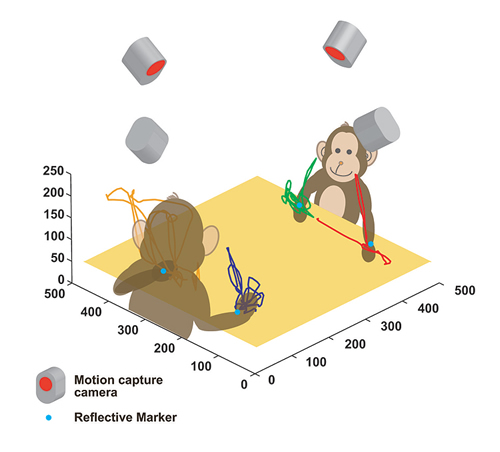
One evolutionary factor of human intelligence is thought to be the acquisition of the ability known as the “social brain”. This means the ability to recognize and actively control social structure in order to achieve one’s wants within the complicated relational structure of human society. Many attempts have been made to scientifically understand the neural mechanism of the “social brain”. However, unlike vision and hearing, it is difficult to create conditions suitable for testing the “social brain” and a neuroscientific approach has yet to be formulated due to the fact that the variable with which the “social brain” deals is “social” structure, something which cannot be described numerically. Thus, we have developed a multi-dimensional recording technique (Fig.1) which allows us to simultaneously record and analyze behavior and brain activity in a new and unprecedented open experimental environment.
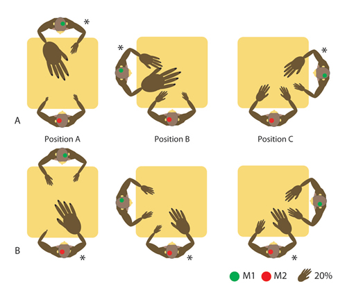
This experiment used two Japanese macaque monkeys (M1, M2), and simultaneously recorded the neuronal activity within the parietal lobe of each. The two monkeys were placed in physical conditions, which were not shared and required no competition (position A) and physical conditions (positions B and C) that were shared. Using motion capture, the behavior of both monkeys as they went about securing food under these conditions was recorded simultaneously together with their neuronal activity.
Behavior analysis observed a clear social pecking order between the two monkeys during task performance, with the subordinate monkey voluntarily refraining from taking food under the shared physical conditions. On the other hand, both monkeys ignored the other and displayed no particular behavioral restraint when physical conditions required no sharing. In other words, this suggests that the social connection between the two was established only after they were forced to share the same space.
During social context-dependent behavior selection, we found a group of parietal cells, that showed motion-related response, altered response characteristics depending on the social conditions (Fig. 2) This indicates that modes of spatial recognition are greatly affected by parietal neurons when social connections exist with others.
This result suggests that spatial recognition within the parietal lobe is not simply a reaction to the physical presence of others but, rather, is a development which occurs only after the presence of others takes on some social meaning. However, the social brain function is not achieved until various other pieces of information, such as the estimation of others’ intentions, wider environmental recognition and further social structure and memory about past experience are unified with behavior recognition of others. In other words, the parietal lobe’s demonstration of social context-dependent neural activity is a part of the network of functions involved in social brain functioning, and in order to gain a detailed understanding social brain function, neural activity from a wider region of the brain will be need to be recorded.
(Naotaka Fujii, Head, Lab. for Adaptive Intelligence)
