


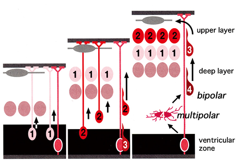
The pyramidal neurons constituting the cerebral cortex are neurons formed from the division of numerous neuronal precursors present in the cerebral ventricular zone of the embryonic cerebrum which then radially migrate toward the superficial layer. The cortex is formed through the radial migration of pyramidal cells adopting an “inside/out pattern” whereby the earliest neurons formed are placed in the deepest layers and later neurons occupy the more superficial layers. The numbers indicate the birth order. During the early part of initial migration there is a stage called “multi-polar” where many neurites are present; afterwards this change to “bipolar” where there are only two processes: one corresponding to a guide process and one to a future axon. After neurons adopt bipolar shape, they migrate in accordance with the structure known as the radial glia.
In a Cdk5-deficient mouse, the morphogenesis of an inside/out pattern shown in Figure 1 is not produced and the position of the II-VI layer is inverted. The abnormalities seen in mutant mice have shown that in a normal cerebral cortex the very process whereby neurons change from being multi-polar to bipolar is the basis of the characteristic formation of neurons seen in the brain.
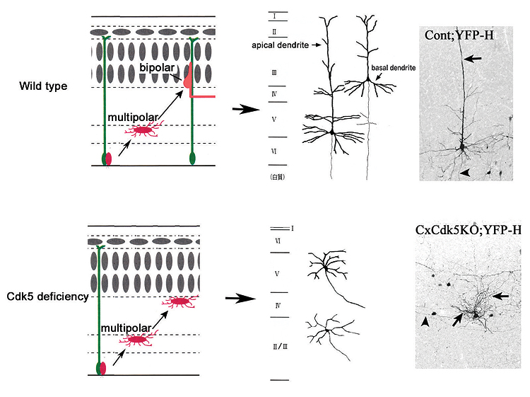
Our brains are extremely delicate, have a well-ordered structure and exhibit surprising abilities. Brains need to be correctly formed in order to function normally. The cerebral cortex of the mammal class has a structure of six regular layers. The structure of these six layers is formed through a skillful mechanism involving the migration of the structure itself from a site of neuronal birth to the site of final placement where it is arranged. At the time of migration the cortex is formed using an “inside/out pattern”, whereby the neurons which developed earliest are distributed in deeper layers while the neurons which developed later are distributed in superficial layers. Recent studies have revealed that, upon migrating, neurons change from being multi-polar, i.e., having many neurites, to bipolar, i.e., having two processes (Fig. 1). In addition, it is known that the “pyramidal neurons” forming the cerebral cortex extend one long dendrite known as an apical dendrite toward the surface of the brain. However, how this form is acquired is not yet clearly understood.
In order to examine the mechanism of neuronal migration and morphological change in neurons, our research group focused on Cyclin-dependent kinase 5 (Cdk5), an enzyme that phosphorylates proteins in neurons. Due to our discovery that the structure of the cerebrum becomes abnormal in mice with Cdk5 deficiencies, we carried out a detailed study of neuronal morphological changes at the time of formation of the cerebral cortex between normal mice and mice without functioning Cdk5 in the cerebral cortex. Our results showed that mice without functioning Cdk5 in their cerebral cortex did not undergo morphological transition from multi-polar to bipolar, and there was little migration from the site of neuronal birth. Furthermore, pyramidal neurons did not extend apical projections to the surface of the brain. This demonstrated that Cdk5 is important to morphological transition in neurons of the cerebral cortex and that neuronal migration is inefficient without the occurrence of morphological transition. We have extrapolated from this result that normal morphological change is responsible for the formation of pyramidal neurons (Fig. 2). While there are several candidates for target proteins requiring phosphorylation of Cdk5, future studies which identify proteins directly involved in morphological transition during the neuronal migration process may provide a more detailed understanding of this mechanism.
